

Structures contributing to virulence
- protein filament “tails” that allow movement and chemotaxis
- Vibrio cholera – 1 flagella
- E. coli – many flagella
- Shigella – no flagella
- Pili (fimbria)
- shorter than flagella and immobile
- allows some bacteria to adhere to their host
- Neisseria
- E. coli
- sex pili are special types of pili used for conjugation rather than virulence
- made of glycoproteins
- Capsules
- prevent phagocytosis
- made of polysaccharide
- except Bacillus anthracis, which contains D-glutamate
- Endospores
- only Bacillus and Clostridium
- dormant form
- no metabolic activity
- confers resistance to hot/cold, wet/dry, and host chemical defenses
- must autoclave to kill
- formed at the end of stationary phase
- when nutrients are in short supply
- dipicolinic acid may confer heat resistance
Other structures
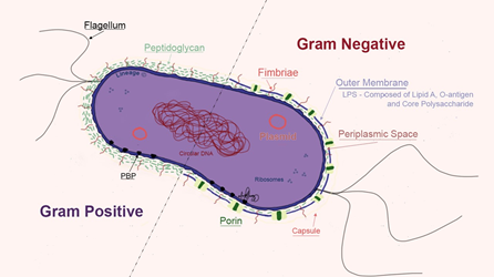
- Cell wall (Gram-positive)
- contains the major surface antigens of Gram-positive bacteria
- e.g. teichoic acid → activates TNF and IL-1
- Mycoplasma has no cell wall
- Mycobacteria has unusual cell wall
- contains the major surface antigens of Gram-positive bacteria
- Outer membrane (Gram-negative)
- source of endotoxin in Gram-negative bacteria
- Peptidoglycan
- structural support
- polymer of sugars and amino acids
- Plasma membrane
- composed of a lipoprotein bilayer
- Ribosome
- 50S and 30S subunits
- protein synthesis
- Plasmid
- DNA that is separate from and replicates independently of chromosomal DNA
- can contain genes for antibiotic resistance and toxins
- Periplasm
- in Gram-negative bacteria, the space between the outer membrane and the cytoplasmic membrane
- contains β-lactamases or other hydrolytic enzymes
- Glycocalyx
- polysaccharide that helps bacteria adhere to host surfaces
- e.g. catheter-associated infection
- polysaccharide that helps bacteria adhere to host surfaces