

Snapshot
- A previously healthy 29-year-old female presents with a progressive, diffuse headache and vomiting. She has no active illnesses, takes a multivitamin, and an oral contraceptive. On exam, there is edema on the scalp, papilledema on fundoscopy, and bilateral muscle weakness. Noncontrast head CT shows a hyperdense lesion in a part of the superior sagittal sinus.
Overview
Introduction
- Structure
- reflections in dura matter where meningeal and periosteal layers split
- Function
- return blood from cerebral veins to internal jugular vein
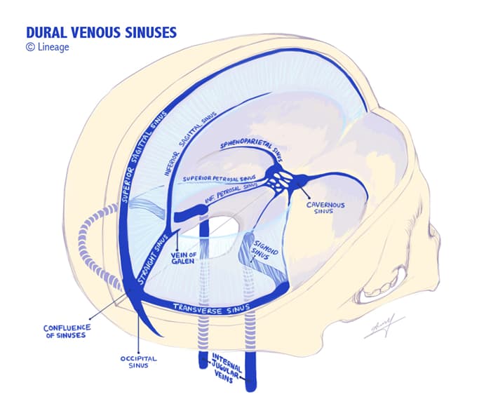
- Main examples
- superior sagittal sinus
- superior to falx cerebri
- tributary of the confluence of sinuses
- inferior sagittal sinus
- inferior to falx cerebri
- tributary to the straight sinus
- cavernous sinus
- lateral to the sella turcica
- tributary of the transverse sinus and sigmoid sinus
- contains CN III, IV, V1, V2, VI, and internal carotid
- clinical correlate
- cavernous sinus thrombosis
- spread of infection from superficial and deep face to cavernous sinus
- present with symptoms relating to compression of cranial nerves
- CN VI usually affected first
- patient cannot abduct eye
- CN VI usually affected first
- cavernous sinus thrombosis
- superior sagittal sinus
- Clinical importance
- venous sinus thrombosis → increases intracranial pressure
- obstruction → increases venous pressure
- consequences include:
- decreases capillary perfusion pressure
- impairs blood brain barrier → vasogenic edema
- impairs CSF reabsorption
- brain parenchymal damage
- venous hemorrhage
- venous sinus thrombosis → increases intracranial pressure