Introduction
- The structures of the eye
- cornea
- a transparent structure that allows light to enter the eye
- pupil
- iris
- covered by the conjunctiva, a transparent mucous membrane
- remember that the conjunctiva lines the inside of the eyelids as well, up to the limbus
- clinical correlate
- conjunctivitis
- which describes inflammation of the conjunctiva
- conjunctivitis
- covered by the conjunctiva, a transparent mucous membrane
- lens
- sclera
- limbus
- which is the border of the cornea and sclera
- medial and lateral canthus
- cornea

- Light enters the eye through the cornea and lens which results in
- an image (inverted and reversed) being formed in the retina
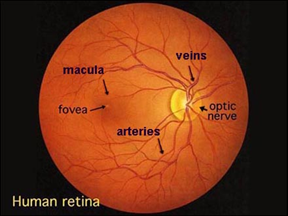
- the area on the retina with the highest visual acuity is the fovea, which is surrounded by the macula
- medial (nasal) to the fovea is the optic disc, which
- is where axons exit forming the optic nerve (cranial nerve II)
- note that the optic nerve does not have photoreceptors over it, resulting in a small blind spot
- is where axons exit forming the optic nerve (cranial nerve II)
- photoreceptors
- there are two classes
- rods
- provides vision in a low-level light environment
- does not detect color
- cones
- highly represented in the fovea
- detect color
- rods
- there are two classes
- an image (inverted and reversed) being formed in the retina
- choroid
- is a vascular layer of the eye
- ciliary body
- is found between the choroid and the iris and is composed of the
- ciliary muscle
- which is controlled by the parasympathetic fibers in the oculomotor nerve in order to
- contract, resulting in miosis
- which is controlled by the parasympathetic fibers in the oculomotor nerve in order to
- ciliary processes
- which have zonular fibers extending from this structure to the lens forming the suspensory ligament
- Lens
- transparent biconvex disc behind the pupil that provides additional refractive power. Is composed of:
- lens capsule
- subcapsular epithelium
- lens fibers
- transparent biconvex disc behind the pupil that provides additional refractive power. Is composed of:
- Anterior chamber
- describes the area behind (posterior) to the cornea and infront (anterior) to the iris
- Posterior chamber
- describes the area posterior to the iris and anterior chamber
- Aqueous humor pathway
- the ciliary body produces aqueous humor into the posterior chamber which
- flows through the space between the lens and iris into the
- anterior chamber and finally drains into the
- trabecular meshwork and then canal of Schlemm
- uveoscleral pathway
- anterior chamber and finally drains into the
- flows through the space between the lens and iris into the
- the ciliary body produces aqueous humor into the posterior chamber which
- Blood supply
- an arterial source is from the ophthalmic artery
- the short posterior, long posterior, and anterior ciliary arteries
- central retinal artery which
- supplies the optic nerve
- venous drainage is from
- the vorticose veins
- central retinal veins
- an arterial source is from the ophthalmic artery


