

Snapshot
- A 27-year-old woman presents to the emergency room with severe hip pain after being a passenger in a head-on motor vehicle accident. On physical exam, she has significant pain and deformity in her left hip. Her left hip is adducted, flexed, and internally rotated.
Introduction
- Clinical definition
- condition in which the femoral head is pushed out of the acetabulum
- in adults, almost always occurs in the setting of significant trauma
- condition in which the femoral head is pushed out of the acetabulum
- Epidemiology
- incidence
- rare injury
- most common mechanism of injury is motor vehicle accident
- 90% of dislocations are posterior
- 10% of dislocations are anterior
- demographics
- 4:1 male-to-female ratio
- most commonly affects adolescents and adults aged 16-40
- risk factors
- significant trauma
- incidence
- Etiology
- traumatic
- developmental
- developmental dysplasia of the hip
- neuromuscular
- cerebral palsy
- Pathoanatomy
- normal anatomy
- hip joint is inherently stable due to
- bony ball-and-socket architecture
- soft tissue constraints
- labrum, joint capsule, and hip musculature
- significant trauma is therefore required to overcome the inherent stability of the joint
- hip joint is inherently stable due to
- mechanism
- axial loading on adducted femur predisposes to posterior dislocation
- dashboard injury
- axial loading on abducted and externally rotated femur predisposes to anterior dislocation
- axial loading on adducted femur predisposes to posterior dislocation
- normal anatomy
- Associated conditions
- 95% incidence of concomitant injuries to other areas of the body
- acetabular and femoral head or neck fractures
- knee ligamentous and meniscal injuries
- closed head injuries
- 95% incidence of concomitant injuries to other areas of the body
- Prognosis
- favorable
- anterior dislocations
- favorable
- simple dislocations (no associated fractures)
Presentation
- Symptoms
- severe pain and immobilty in the affected hip
- may also complain of lower back, thigh, knee, or lower leg pain
- Physical exam
- hip position
- anterior dislocation
- hip will be flexed, abducted, and externally rotated
- anterior dislocation
- pain with passive or active movement
- hip position
- thorough head-to-toe examination following Advanced Trauma Life Support (ATLS) protocols must be performed given high incidence of concomitant head and extremity injuries
Imaging
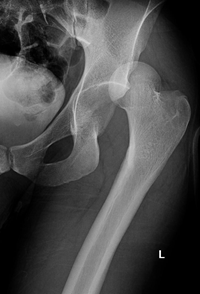
- Radiographs
- indication
- anteroposterior (AP) pelvis radiograph always indicated when hip dislocation is suspected
- indication
- Computerized tomography (CT) scan
- indication
- high suspicion for associated fractures
- finding
- indication
- associated fractures to acetabulum, femoral head, and femoral neck
Differential
- Femoral neck fracture
- hip will remain in acetabulum on AP pelvis radiograph
- Acetabular fracture
- hip will remain in acetabulum on AP pelvis radiograph
Treatment
- Conservative
- closed reduction under conscious sedation
- indication
- closed reduction should be attempted in all traumatically dislocated hips
- indication
- closed reduction under conscious sedation
- Operative
- open reduction
- indication
- open reduction
- failure of closed reduction
Complications