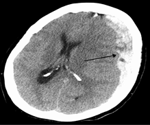
| Intracranial Hemorrhage |
| Type | Pathogenesis | Presentation and Management | Head CT |
Epidural hematoma
| Typically secondary to rupture of the middle meningeal artery in the setting offracture of the temporal bone (pterion)Recall that the middle meningeal artery is a branch off of themaxillary artery | Initially there may be no symptoms (lucid interval) temporal bone fracture may present with hearing loss, periauricular ecchymosis, facial paralysis, hemotympanum, and/or dizziness As the hematoma grows, itleads to brain tissue compression whichincreases intracranial pressureThis increased intracranial pressure can result inbrain herniation such astranstentorial herniation Managementcraniotomy and hematoma evacuation when indicated | Lens-shaped biconvexhematoma secondary to a rapidly expanding hematoma that peels the dura away from the skullrecall that this is due to being under arterial pressure |
| Subdural hematoma | Secondary to rupture of thebridging veinsThe most common cause ishead trauma (e.g., falls, assaults, and motor vehicle accidents)Risk factorssignificant cerebral atrophy, such as inthe elderlychronic alcohol abuseprevious traumatic brain injury | Clinical presentation depends on if it is achronic subdural hematomaacute subdural hematomaChronic subdural hematomatypically seen in the elderlycan be seen with minimal or absent history of head traumavague symptoms such asheadachecognitive impairmentunsteady gaitthe focal accumulation of blood can result infocal seizuresfocal neurologic deficitsAcute subdural hematomatypically has a history of traumatic injurysymptoms of increased intracranial pressure such asheadachevomitingcranial nerve palsiesManagementsurgical removal (e.g., craniotomy or burr hole) | Blood accumulates between the dura and the arachnoid which createsa crescent shaped hematoma |
| Subarachnoid hemorrhage | Most commonly due to arterial aneurysm rupture in the subarachnoid space, which can result from traumatic causes non-traumatic causes (spontaneous rupture)Less commonly due toarteriovenous malformationRisk factorsatherosclerotic diseasesmokingexcessive alcohol intakepolycystic kidney diseaseEhlers-Danlos syndromefibromuscular dysplasia | Sudden “thunderclap” headache or”the worst headache of my life”Meningeal irritationphotophobianuchal rigidityCan also result infocal neurologic deficitsimpaired conciousnesscomaManagementevaluate all cerebral vessels for aneurysm location(e.g., angiogram)oral or via nasograstric tube nimodipine should be administered to prevent cerebral vasospasm however, it does not angiographically improve vasospasmimprove outcomessurgical clipping orendovascular coiling | Blood accumulates within the subarachnoid space, where the major blood vessels of the brain are housedBlood can be found around the sulci and contours the pia A non-contrast head CT is used and will detect blood ifperformed within the first 3 days after aneurysm ruptureNote a lumbar puncture should be performed ifthe non-contrast head CT is negative andclinical suspicion for subarachnoid hemorrhage is high |
| Hypertensive hemorrhage | Secondary to uncontrolled hypertension (HTN)note that HTN is a common cause of intracerebral hemorrhagesHTN on the small vessels can result inlipohyalinosisCharcot-Bouchard microaneurysms | Symptoms depend on where the hemorrhage occursfor example, putamenal hemorrhages can result inhemiplegiahemisensory lossgaze palsycomahow large the hemorrhage isif large, it can result insymptoms of increased intracranial pressureManagementinvolves both medical and surgical interventions | Hyperintense lesions can be seen on non-contrast head CT (just like in other causes of an acute bleed) in typical locations such as basal ganglia (most common)thalamuscerebellumpons |
| Lobar hemorrhage | Can be secondary toamyloid angiopathy (most common cause)seen in older patients (> 50 years of age)HTNAmyloid can deposit in the vessel wall, makingit fragile and thusprone to bleed | Symptoms depend on where the hemorrhage is such asparietal lobeoccipital lobee.g., contralateral homonymous hemianopsiahow large the hemorrhage isif large, it can result insymptoms of increased intracranial pressurethese patients are at higher risk for seizures than hypertensive hemorrhagesManagementinvolves both medical and surgical interventions | Hyperdense lesion affecting a particular lobe (e.g., parietal and occipital) on non-contrast head CT |


