

Overview Kidney Embryology
Kidney development
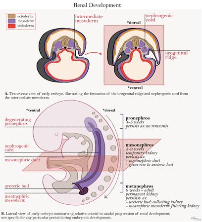
- progresses in a cranial to caudal direction
- intermediate mesoderm → urogenital ridge → nephrogenic cord → urinary system
- Pronephros
- non-functional
- appears by week 4
- degenerates by week 5
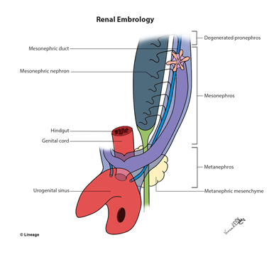
- Mesonephros
- development induced by pronephric duct
- forms mesonephric duct (Wolffian duct)
- interim kidney for 1st trimester
- opens into urogenital system and gives rise to male genital system
- Metanephros
- develops from mesonephric outgrowth called ureteric bud during week 5
- fully canalized and functioning at week 10
- nephrogenesis continues through 32 – 36 weeks of gestation
- derivatives include
- Ureteropelvic junction with kidney
- Adult kidney
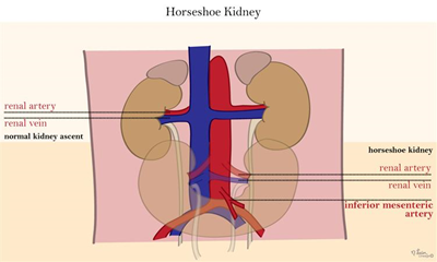
- embryo grows faster caudally causing a change in location of the kidney from S1 – S2 to a final position of T12 – L3
Developmental Abnormalities
- Renal agenesis
- failure of ureteric buds to form → no kidney formation
- Potter’s Syndrome
- Horseshoe kidney
normal kidney function
Introduction
The kidneys are essential organs responsible for regulating fluid balance, electrolyte levels, and waste excretion in the human body. Understanding the embryology of the kidneys is vital for medical students preparing for the USMLE exam, as it provides a foundation for comprehending their anatomy, development, functions, and associated clinical considerations. This article aims to provide a comprehensive overview of kidney embryology, types, treatment, and related studies.
Embryology of the Kidneys
The kidneys originate from intermediate mesoderm during the fifth week of embryonic development. The process begins with the formation of the pronephros, a transient structure that quickly regresses and is non-functional in humans. Subsequently, the mesonephros develops, which consists of mesonephric tubules that contribute to the formation of the male reproductive system.
Eventually, the metanephros arises as a definitive kidney, forming from the ureteric bud and metanephric mesenchyme. The ureteric bud grows into the metanephric mesenchyme, inducing its differentiation into nephrons and the collecting system. These complex interactions result in the formation of the renal cortex, medulla, and collecting ducts.
Treatment Considerations and Associated Studies
Kidney diseases, such as chronic kidney disease, kidney stones, and renal tumors, require careful management. Treatment options range from medication and lifestyle modifications to surgical interventions, dialysis, and kidney transplantation. Diagnostic studies used to assess kidney disorders and determine appropriate treatment strategies include renal ultrasound, computed tomography (CT) scan, magnetic resonance imaging (MRI), renal biopsy, and blood tests to evaluate renal function, electrolyte levels, and urine composition.
Types of Nephrons
Nephrons are the functional units of the kidneys responsible for filtration, reabsorption, and secretion. Two types of nephrons exist: cortical nephrons and juxtamedullary nephrons. Cortical nephrons, which make up approximately 85% of all nephrons, have their glomeruli located in the outer cortex. Juxtamedullary nephrons, on the other hand, have glomeruli located near the corticomedullary junction and play a crucial role in establishing the concentration gradient necessary for urine concentration.
Renal Developmental Anomalies
During kidney development, various anomalies can occur, leading to structural or functional abnormalities. Examples of renal developmental anomalies include renal agenesis (complete absence of one or both kidneys), renal hypoplasia (underdevelopment of one or both kidneys), renal dysplasia (abnormal tissue formation within the kidneys), and polycystic kidney disease (formation of fluid-filled cysts within the kidneys). These anomalies may result in renal dysfunction, requiring specialized management approaches tailored to the specific condition.
Conclusion
A thorough understanding of kidney embryology is essential for medical students preparing for the USMLE exam. Knowledge of kidney development aids in comprehending the complex anatomy and physiological functions of the kidneys. Additionally, familiarity with associated clinical conditions, diagnostic studies, and treatment options is vital for diagnosing and managing kidney disorders effectively.
Check out Ultimate USMLE Step 1 Study Notes.