Snapshot

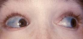
- A 4-year-old girl falls down on the playground and is rushed to the emergency room. Her work-up reveals a new fracture in her right tibia, as well as multiple old fractures in her bilateral arms and legs at various stages of healing. Her physician is concerned about child abuse, but communication with the girl’s pediatrician reaveals her condition. On exam, her sclera are blue.
Introduction
- Defect in production of type I collagen causing abnormal, fragile bones that fracture easily
- Most commonly autosomal dominant
- also autosomal recessive
- Also known as “brittle bone disease”
- Pathogenesis
- type I collagen = most common type (90% of all)
- found in bone, skin, tendon, dentin, cornea, wound repair, fascia
- result from
- defect in forming triple helix (procollagen) = abnormal collagen
- defective glycosylation of hydroxylysine residues
- ↓ production of type I collagen
- defect in forming triple helix (procollagen) = abnormal collagen
- type I collagen = most common type (90% of all)
- Often confused with child abuse
Presentation
- Symptoms
- recurrent fractures with minimal force
- often during birth
- recurrent fractures with minimal force
- Physical exam
- dental abnormalities (brown, opalescent teeth)
Evaluation
- Diagnosis made by clinical and radiographic findings (skeletal survey)
- Confirmed with DNA (blood or saliva) or protein testing (skin biopsy)
Differential
- Non-accidental injury
- Idiopathic juvenile osteoporosis
Treatment
- Bisphosphonates to increase bone mineral density
Prognosis, Prevention, and Complications
- Prognosis
- recurrent fractures
- no cure for disease
- Prevention
- screening for mutation in family members
- prenatal screening with chorionic villus sampling
- Complications
- recurrent fractures leads to deformity and chronic pain
- may result in wheelchair dependence
- hearing loss


