

Snapshot
- A 10-year-old boy is brought to his dermatologist for a developing rash. A couple of weeks ago, he recovered from a common cold. A week after, he developed an oval rash on his chest. Thinking it was a fungus infection, his parents applied anti-fungal cream to the area. However, a week after the first lesion appeared, he developed multiple smaller rashes in his lower abdomen. They are sometimes itchy, but only mildly so.
Introduction
- Common, self-limited papulosquamous eruption
- Pathogenesis
- idiopathic
- often associated with URI
- seasonal pattern suggests viral etiology, though not confirmed
- potential link to herpesvirus types 6 and 7
- Epidemiology
- children
- young adults
Presentation
- Symptoms
- prodrome or URI within a month of onset
- little or no pruritus
- Physical exam
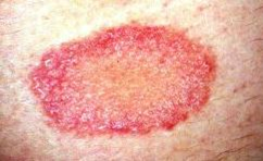
- herald patch, a single lesion
- usually on the trunk
- plaque with thin collarette of scale inside the border
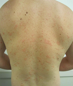
- eruption in 1-2 weeks
- multiple smaller papules appear in “Christmas tree” distribution
- oriented along Langer (skin cleavage) lines
- rose-colored or violet
- multiple smaller papules appear in “Christmas tree” distribution
- resolution in 4-12 weeks
- herald patch, a single lesion
- resolves spontaneously without scarring
Evaluation
- Diagnosis from clinical exam and history
- Diagnosis confirmed with skin biopsy
- potassium hydroxide preparation to exclude Tinea spp.
Differential Diagnosis
- Tinea corporis
- Secondary syphilis (especially if palm and soles involved)
- Tinea versicolor
- Drug eruption
- Guttate psoriasis
Treatment
- Observation
- lesions heal within 4-12 weeks
- Natural sunlight
Prognosis, Prevention, and Complications
- Prognosis
- very good
- typically self-limited and self-resolving in 4-12 weeks
- Complications
- relapse