

Overview
Snapshot
- A 13-year-old boy with cystic fibrosis presents to the emergency department with S. aureus pneumonia. He suddenly develops increased respiratory distress, pleuritic chest pain, and decreased breath sounds in the left chest.
Introduction
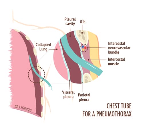
- Pneumothorax results from air trapped in the pleural space that collapses a portion of the ipsilateral lung
- Etiologies
- primary spontaneous
- caused by rupture of the subpleural apical blebs
- usually occurs in tall, thin males
- secondary spontaneous
- spontaneous pneumothorax in the setting of underlying lung disease (ie COPD, TB, trauma, pneumocystis pneumonia, S. aureus)
- primary spontaneous
- subclavian access has the highest risk of pneumothorax due to the proximity to the apices of the lungs
Presentation
- Symptoms
- sudden-onset, unilateral, pleuritic chest pain
- dyspnea
- acute respiratory distress
- Physical exam
- decreased or absent breath sounds
- hyperresonant to percussion
- decreased or absent tactile fremitus
- may see tracheal deviation if tension pneumothorax present
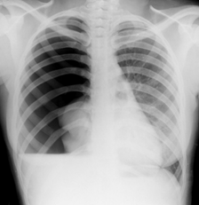
Evaluation
- CXR
- diagnostic gold standard in upright patient