

Overview
Snapshot
- A 22-year-old man presents to the emergency department after being involved in a motor vehicle accident. The patient is conscious but reports knee pain. He was wearing a seat belt, denies any head trauma, but says that his knees collided with the dashboard. Physical examination is significant for increased laxity of the proximal tibia with posterior force. A magnetic resonance imaging of the knee is consistent with a posterior cruciate ligament tear.
Introduction
- Clinical definition
- injury of the posterior cruciate ligament (PCL)
- Epidemiology
- risk factors
- high-energy trauma (e.g., motor vehicle accident)
- sport activities
- risk factors
- Etiology
- trauma that posteriorly translates the proximal tibia
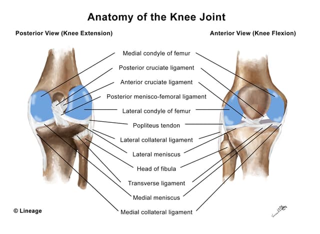
- Pathoanatomy
- normal anatomy
- the largest intraarticular ligament in the knee
- originates from the lateral portion of the medial femoral condyle and inserts at the fovea centralis (PCL facet)
- pathology
- posteriorly directed shear force when the knee is flexed
- “dashboard injury” (high-energy trauma)
- when the proximal tibia collides with the dashboard of a car in a motor vehicle accident
- sports injuries (low-energy trauma)
- the player falls on their knee while it is flexed and the foot is plantarflexed
- direct trauma to the anterior knee
- “dashboard injury” (high-energy trauma)
- posteriorly directed shear force when the knee is flexed
- normal anatomy
- Associated conditions
- high-energy trauma can result in PCL injury as well as injury to the
- posterolateral corner
- anterior cruciate ligament
- medial collateral ligament
- high-energy trauma can result in PCL injury as well as injury to the
- Prognosis
- insufficient evidence to determine the true prognosis
Presentation
- Symptoms
- may present with posterior knee pain
- athletes may continue playing sports and not seek medical care
- may present with posterior knee pain
- Physical exam
- posterior drawer test
- the proximal tibial is pushed posteriorly to assess for PCL laxity while the knee is flexed at 90 degrees
- posterior translation is considered a positive test
- the proximal tibial is pushed posteriorly to assess for PCL laxity while the knee is flexed at 90 degrees
- mild-to-moderate effusion
- slight limp or antalgic gait
- posterior drawer test
- may have impaired terminal knee flexion
Imaging
- Radiography
- indication
- performed initially to assess for fractures
- indication
- Magnetic resonance imaging (MRI)
- indication
- typically performed in all patients presenting with acute PCL injury
- indication
- highly sensitive and accurate for acute PCL injury
Studies
- Making the diagnosis
- a presumptive diagnosis can be made based on clinical presentation
- MRI can definitively diagnose acute or subacute PCL injury
Differential
- Anterior cruciate ligament injury
- distinguishing factor
- patients typically hear a “pop” after injury and there is typically significant edema and joint effusion
- distinguishing factor
- patients have a positive anterior drawer test
Treatment
- Conservative
- rest, ice, compression, and elevation (RICE) therapy
- indication
- standard management of patients with an isolated PCL injury
- in addition to RICE therapy, patients should receive
- nonsteroidal anti-inflammatory drugs (NSAIDs) or acetaminophen for pain management
- immobilization in extension (e.g., hinged knee brace)
- in addition to RICE therapy, patients should receive
- standard management of patients with an isolated PCL injury
- indication
- rest, ice, compression, and elevation (RICE) therapy
- Operative
- reconstructive surgery
- indication
- reconstructive surgery
- typically reserved for acute and multi-ligamentous knee injuries
Complications
- May result in osteoarthritis