

Fetal-postnatal derivatives
Aortic arch derivatives
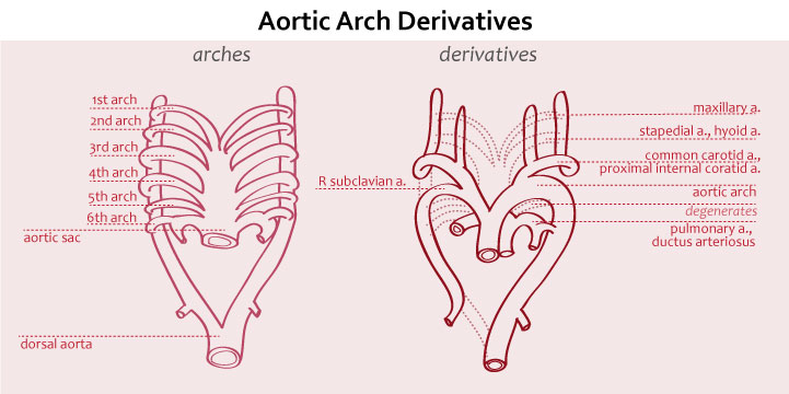
| Arch | Derivative |
| 1st | Part of maxillary artery branching from external carotid |
| 2nd | Stapedial artery and hyoid artery |
| 3rd | Common carotid arteries and proximal part of internal carotid arteries |
| 4th | On left, aortic archOn right, proximal part of right subclavian artery |
| 5th | Degenerates during development |
| 6th | Proximal part of pulmonary arteriesDuctus arteriosus |
In medicine, “postnatal derivatives” refer to anatomical structures or physiological functions that undergo significant changes or adaptations after birth. The understanding of these changes is crucial for healthcare professionals, particularly in neonatology and pediatric medicine. This article provides a comprehensive overview of postnatal derivatives, including their types, functions, studies, and treatments.
Types of Postnatal Derivatives:
- Ductus Arteriosus (DA): The DA is a blood vessel connecting the pulmonary artery and the aorta in fetal circulation. After birth, it should naturally close, becoming the ligamentum arteriosum. However, in some cases, it remains open, leading to a condition known as Patent Ductus Arteriosus (PDA).
- Foramen Ovale (FO): During fetal life, the FO is a hole between the two atria that allows blood to bypass the non-functional fetal lungs. Once the baby starts breathing after birth, the FO should close and eventually become the fossa ovalis. Failure of closure results in a Patent Foramen Ovale (PFO).
- Ductus Venosus (DV): The DV shunts oxygenated blood from the umbilical vein to the inferior vena cava in the fetus. After birth, it becomes the ligamentum venosum.
- Umbilical Arteries and Vein: The umbilical cord contains two arteries and one vein, which supply nutrients and oxygen to the fetus. After birth, they become fibrotic cords.
Functions and Significance:
The postnatal closure or adaptation of these derivatives is vital for a successful transition from intrauterine to extrauterine life. The closure of the DA and FO allows the baby’s circulatory system to function independently of the placenta. The establishment of the independent pulmonary and systemic circulation is critical for adequate oxygenation of organs and tissues.
Clinical Studies and Abnormalities:
Research on postnatal derivatives and their abnormalities is an essential aspect of neonatal and pediatric medicine. Several diagnostic methods help identify any issues:
- Echocardiography: This non-invasive imaging technique uses sound waves to visualize the heart’s structure and function. It helps detect conditions like PDA, PFO, and other congenital heart defects.
- Doppler Studies: Doppler ultrasound assesses blood flow patterns in vessels, aiding in the evaluation of DV functionality and potential abnormalities.
- X-rays and MRI: These imaging techniques provide detailed images of the heart and surrounding structures, aiding in the diagnosis of structural anomalies.
Treatment and Management:
The management of postnatal derivatives varies depending on the specific condition and its severity:
- Conservative Management: In some cases, postnatal derivatives may close on their own without intervention, especially if they are small and clinically insignificant.
- Medication: Pharmacological agents, such as nonsteroidal anti-inflammatory drugs (NSAIDs), can be used to promote the closure of a PDA.
- Surgical Intervention: Persistent and significant postnatal derivatives may require surgical correction. For example, large PDAs or certain congenital heart defects might necessitate surgical closure.
- Catheter-Based Procedures: Minimally invasive catheter-based interventions can be used to close certain congenital heart defects, offering an alternative to open-heart surgery.
Complications and Prognosis:
If left untreated, some postnatal derivatives can lead to complications:
- Congestive Heart Failure (CHF): In cases of a large, untreated PDA, increased blood flow to the lungs can strain the heart and lead to CHF.
- Risk of Thromboembolism: A PFO can act as a pathway for blood clots to travel from the venous circulation to the arterial circulation, potentially causing strokes or other thromboembolic events.
Conclusion:
Postnatal derivatives play a significant role in the transition from fetal to neonatal circulation. Understanding their closure and adaptation is essential for healthcare professionals dealing with newborns and infants. Timely recognition and appropriate management of abnormalities can significantly impact the prognosis and overall health of neonates.
Advances in diagnostic techniques and treatment modalities continue to improve outcomes and reduce the complications associated with these conditions, making the study of postnatal derivatives a crucial aspect of medical education and practice.
Check out USMLE Success Strategy: Personalized Consultation and Study Plan Development.