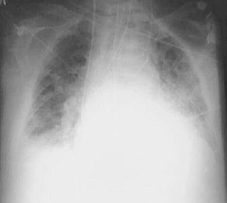
Introduction – Pulmonary Edema

- Fluid accumulation in air spaces and parenchyma of the lungs
- Pathophysiology
- edema arises due to an imbalance in hydrostatic and/or oncotic pressure
- increased hydrostatic pressure in the pulmonary capillaries (Pc)
- cardiogenic causes (see below)
- decreased oncotic pressure in the pulmonary capillaries (πc)
- kidney or liver dysfunction (e.g. nephrotic syndrome, cirrhosis)
- movement of fluid is driven by Starling forces
- increased hydrostatic pressure in the pulmonary capillaries (Pc)
- edema arises due to an imbalance in hydrostatic and/or oncotic pressure
- Causes
- changes in pressure are of cardiogenic or non-cardiogenic origin
- cardiogenic causes include
- heart failure
- left heart failure
- volume overload
- mitral stenosis
- in all cases above, an increase in left ventricular end diastolic volume increases hydrostatic pressure in LV, LA, pulmonary vein, and pulmonary capillaries
- the increase in hydrostatic pressure overwhelms the oncotic pressure in the pulmonary capillaries
- a transudate leaks into the interstitial space and ultimately into the alveoli
- heart failure
- non-cardiogenic causes include
- infection
- sepsis
- pneumonia
- aspiration
- drowning
- gastric aspiration
- drugs
- heroin
- high altitude
- ARDS
- infection
- alveolar-capillary damage
Overview
Pulmonary edema is a medical condition characterized by the accumulation of fluid in the lungs. The excess fluid buildup in the lungs can make it difficult to breathe and can lead to a range of symptoms, including shortness of breath, coughing, and wheezing.
There are two main types of pulmonary edema: cardiogenic pulmonary edema and non-cardiogenic pulmonary edema. Cardiogenic pulmonary edema occurs as a result of heart failure, where the heart is unable to pump blood effectively, causing blood to back up into the lungs. Non-cardiogenic pulmonary edema, on the other hand, can be caused by a variety of factors, including severe infections, exposure to toxic chemicals, or damage to the lungs from trauma or injury.
Presentation
- Symptoms
- dyspnea
- including orthopnea and paroxysymal nocturnal dyspnea (PND)
- dyspnea
- Physical exam
- bibasilar inspiratory crackles
- due to air expanding fluid-filled alveoli
- rusty-colored sputum
- due to rupture of pulmonary capillaries from elevated hydrostatic pressure
- wheezing
- due to peribronchiolar edema
- bibasilar inspiratory crackles
- “cardiac asthma”
Evaluation
- CXR
- congestion in upper lobes
- perihilar congestion
- “bat wing configuration”
- alveolar infiltrates
- Kerley’s lines
- due to septal edema
- Biopsy
- macrophages phagocytose blood following rupture of capillaries
Treatment
The treatment of pulmonary edema depends on the underlying cause of the condition. In general, the goal of treatment is to remove the excess fluid from the lungs and improve breathing.
If pulmonary edema is caused by heart failure, treatment may include:
- Diuretics: These medications help remove excess fluid from the body and reduce the workload on the heart.
- Vasodilators: These medications help dilate the blood vessels and reduce the workload on the heart.
- Inotropes: These medications help improve the heart’s pumping ability.
- Oxygen therapy: This can help increase oxygen levels in the blood and reduce breathing difficulties.
- Mechanical ventilation: In severe cases, a person may require a breathing machine to help them breathe.
If pulmonary edema is caused by a lung infection or other non-cardiac condition, treatment may include:
- Antibiotics: These medications can help treat the infection and reduce inflammation in the lungs.
- Corticosteroids: These medications can help reduce inflammation in the lungs.
- Oxygen therapy: This can help increase oxygen levels in the blood and reduce breathing difficulties.
- Mechanical ventilation: In severe cases, a person may require a breathing machine to help them breathe.
It’s important to seek medical attention if you are experiencing symptoms of pulmonary edema. Prompt treatment can help improve outcomes and prevent complications.
Studies
There have been numerous studies conducted on pulmonary edema, exploring its causes, risk factors, diagnosis, and treatment. Here are a few examples of recent studies:
- A study published in the Journal of Critical Care in 2021 found that a combination of diuretics and nitrates was effective in reducing mortality in patients with acute pulmonary edema.
- A study published in the American Journal of Respiratory and Critical Care Medicine in 2020 found that a machine learning model could accurately predict the development of pulmonary edema in patients with heart failure.
- A study published in the Journal of the American College of Cardiology in 2019 found that the use of non-invasive ventilation in patients with acute cardiogenic pulmonary edema was associated with reduced mortality and fewer complications.
- A study published in the Journal of Thoracic Imaging in 2018 found that computed tomography (CT) imaging could help differentiate between cardiogenic and non-cardiogenic pulmonary edema, which could aid in diagnosis and treatment.
- A study published in the Journal of the American Medical Association in 2017 found that early administration of high-dose furosemide (a diuretic) was associated with improved outcomes in patients with acute pulmonary edema.
Check out USMLE Step 1 Mastery: Comprehensive Course and Lecture Notes.


