Snapshot

- A 50-year-old woman presents to her physician’s office for a routine checkup. Once at the office, she reports that she has been generally doing well but recently noticed that her fingers tend to turn blue in the cold. She reports feeling a general skin tightening in her face and hands, which makes forming a fist difficult. She also notes that she has had increased acid reflux lately and requests a medication for that. Her past medical history includes autoimmune thyroid disease and alopecia areata. Physical exam reveals sclerodactyly, and tight, hardened skin with limited mobility in her fingers. Her physician sends her for additional autoimmune workup.
Introduction
- Clinical definition
- an autoimmune skin disease characterized by progressive hardening and induration of the skin and/or other structures such as the subcutaneous tissues, muscles, and internal organs
- triad
- autoimmunity
- noninflammatory vasculopathy
- collagen deposition with fibrosis
- triad
- classification
- localized scleroderma
- affecting only the skin and muscles
- systemic scleroderma (systemic sclerosis)
- affecting internal organs including the kidney, lung, and heart
- further subdivided into limited and diffuse
- limited progresses more slowly and has less internal organ involvement
- subtype of systemic scleroderma is CREST syndrome
- Calcinosis cutis
- anti-Centromere antibody
- Raynaud phenomenon
- ↓ blood flow to skin from either cold temperatures or stress, which causes vasospasms
- colors of affected area, commonly the digits, change from white (ischemia) to blue (hypoxia) to red (re-perfusion)
- Esophageal dysmotility
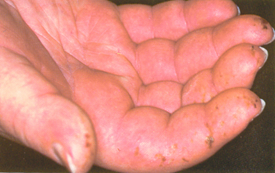
- Sclerodactyly
- Telangiectasia
- localized scleroderma
- an autoimmune skin disease characterized by progressive hardening and induration of the skin and/or other structures such as the subcutaneous tissues, muscles, and internal organs
- Epidemiology
- demographics
- female > male
- African Americans > Caucasian
- 30-50 years of age but can affect all ages
- risk factors
- exposure to potential triggers
- demographics
- Etiology
- multifactorial, including genetic predisposition and environmental triggers
- possible triggers include silica, solvent (such as benzene), and radiation exposure
- multifactorial, including genetic predisposition and environmental triggers
- Pathogenesis
- sclerosis
- excessive deposition of collagen and other elements of the extracellular matrix in skin and internal organs
- fibroproliferation of microvasculature leading to a noninflammatory vasculopathy
- chronic inflammation with alterations of humoral and cellular immunity
- increased release of inflammatory cells help initiate and propagate the fibrotic process
- esophageal dysmotility
- atrophy of smooth muscles in esophagus can cause ↓ lower esophageal sphincter pressure and dysmotility, leading to increased dysphagia and acid reflux
- sclerosis
- Associated conditions
- other autoimmune diseases
- Prognosis
- limited scleroderma is more benign
Presentation
- Symptoms
- skin
- diffuse pruritus
- gastrointestinal
- acid reflux
- respiratory
- progressive dyspnea
- dry cough due to restrictive lung disease
- musculoskeletal
- myalgias
- arthralgias
- cardiac
- palpitations or irregular heart beats
- skin
- Physical exam
- skin
- skin tightness, induration, and hardening
- affecting the fingers (sclerodactyly)
- shiny with loss of “wrinkles” from skin folds
- limited mobility due to skin tightening
- digital ulceration
- edema not responsive to diuresis
- hyper and hypopigmentation
- telangiectasias on skin and mucosa
- skin tightness, induration, and hardening
- respiratory
- dry rales indicative of pulmonary involvement
- cardiac
- symptoms of cor pulmonale if there is pulmonary involvement
- jugular venous distention
- edema
- hepatomegaly
- symptoms of cor pulmonale if there is pulmonary involvement
- renal
- skin
- hypertension
Imaging
- Computerized tomography (CT) scan
- indications
- evaluate pulmonary involvement
- view
- chest
- findings
- ground-glass appearance may indicate early lung fibrosis
- indications
- honeycombing and bronchiolectasis indicate developed interstitial fibrosis
Evaluation
- Labs
- anti-centromere autoantibody
- associated with limited scleroderma (CREST syndrome)
- in ~ 50% of patients
- antinuclear antibodies
- in ~ 90-95% of affected patients
- speckled or centromere pattern
- nucleolar pattern is specific for systemic sclerosis
- ↑ inflammatory markers
- erythrocyte sedimentation rate
- C-reactive protein
- serum creatinine
- to monitor for renal involvement
- ↑ CXCL4
- may indicate pulmonary fibrosis
- ↑ N-terminal probrain natriuretic peptide
- may indicate early pulmonary hypertension
- anti-centromere autoantibody
- Electrodiagnostics
- routine EKG to assess for cardiac involvement
- Pulmonary function test
- to detect early signs of pulmonary fibrosis
- Making the diagnosis
- based on clinical presentation and laboratory studies
Differential
Treatment
- Management approach
- largely based on symptomatic relief
- Medical
- immunosuppressive therapies
- indication
- to prevent progression of sclerosis, especially if pulmonary system is involved
- drugs
- methotrexate
- mycophenolate mofetil
- cyclophosphamide
- reserved for when disease is refractory to either methotrexate of mycophenolate mofetil
- indication
- angiotensin-converting enzyme (ACE) inhibitor
- indication
- renal involvement of systemic sclerosis
- indication
- anti-histamines
- indication
- pruritus
- indication
- ambrisentan (endothelin receptor antagonist) and tadalafil (phosphodiesterase type 5 inhibitor) combination therapy
- indication
- immunosuppressive therapies
- pulmonary hypertension
Complications


