

Snapshot
- A 40-year-old woman with no significant past medical history is bothered by several brown moles. She started noticing them in the past year and notes that they have not changed in size. There is one mole in particular that is bothersome to her, cosmetically, because it is large. She denies any family history of skin cancer, but recalls seeing similar moles on her mother. She opts to use cryotherapy to remove the lesions.
Introduction
- Common, benign persistent epidermal proliferations with variable appearances
- Can mimic malignancies, especially melanoma
- Genetics
- can be inherited
- Epidemiology
- rare before 30 years old
- one of the most common benign growths
- Associated conditions
- underlying GI or lymphyoid malignancies
- NOT related to actinic keratosis or seborrheic dermatitis (despite the similarity in name)
- Prognosis
- no risk for progression to malignancies
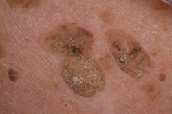
Presentation
- Symptoms
- patients can often scratch off a lesion
- Physical exam
- usually multiple lesions
- variable appearance
- flat or raised
- smooth, velvety, or verrucous
- color ranges from white, pink, brown, or black
- even within a single lesion, color may vary
- common on trunk, face, extremities
Evaluation
- If clinically mimicking skin cancer (e.g., with very dark pigmentation suspicious of melanoma)
- skin biopsy
Differential
- Melanoma
- can be mistaken for melanoma
Treatment
- Medical
- observation only
- indications
- most cases are treated with observation alone
- indications
- liquid nitrogen (cryotherapy)
- indications
- cosmetic concerns
- indications
- observation only