

Introduction
- Secondary lymphoid tissue has many important roles in immunity including
- filtration and sampling of bodily fluids in order to detect infection
- sequestration of pathogens and ingestion by innate immune cells
- activation of adaptive immunity by antigen presenting cells
- maintainance of immunological memory by storage of long lived cells
- There are several types of secondary lymphoid tissue including the
- spleen
- a solid organ in the left upper quadrant of the abdomen
- lymph nodes
- a diffuse network of small nodes throughout the body
- mucosal lymphoid tissue
- patches of immunological tissue such as
- Waldeyer’s ring consisting of
- tubular tonsils in the pharyngeal recess
- palatine tonsils
- lingual tonsils at the base of the tongue
- Waldeyer’s ring consisting of
- patches of immunological tissue such as
- spleen
- Peyer patches in the lower jejunum and ileum
Spleen
- The spleen is located in the left upper quadrant of the abdomen where
- it is protected by the 9th to 11th ribs
- it sits anterior to the left kidney
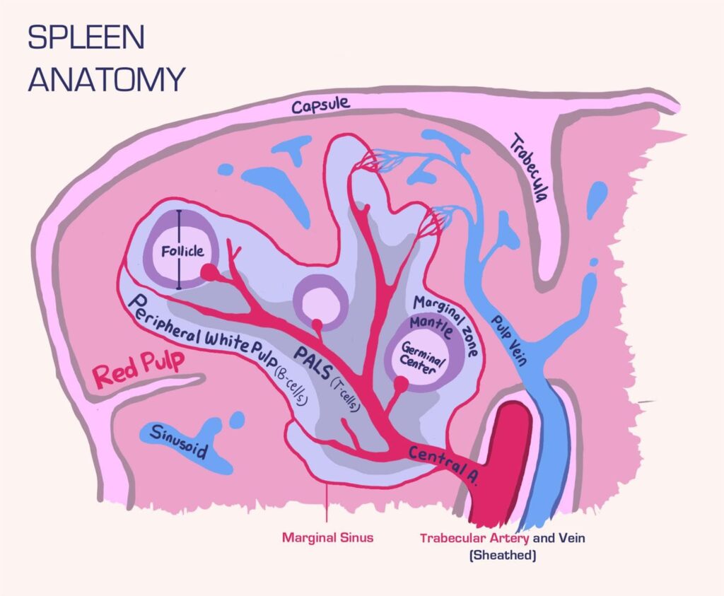
- There are several components of the spleen, which are divided into
- white pulp, which is the
- the immunologically active part of the spleen containing
- germinal centers containing
- activated B-cells (part of a larger B-cell follicle)
- periarteriolar lymphatic sheaths (PALS) containing
- T-cells
- marginal zones containing
- macrophages and other antigen-presenting cells
- germinal centers containing
- the immunologically active part of the spleen containing
- red pulp, which is
- white pulp, which is the
- There are many clinical manifestations of asplenia including
- increased susceptibility to infections by encapsulated bacteria
- appearance of abnormal RBC forms on peripheral blood smear
- others described more fully in the asplenia topic
Lymph Nodes
- Lymph nodes are a diffuse network of encapsulated lymphoid tissue that
- have many regional afferent lymphatic vessels
- have at least one efferent lymphatic
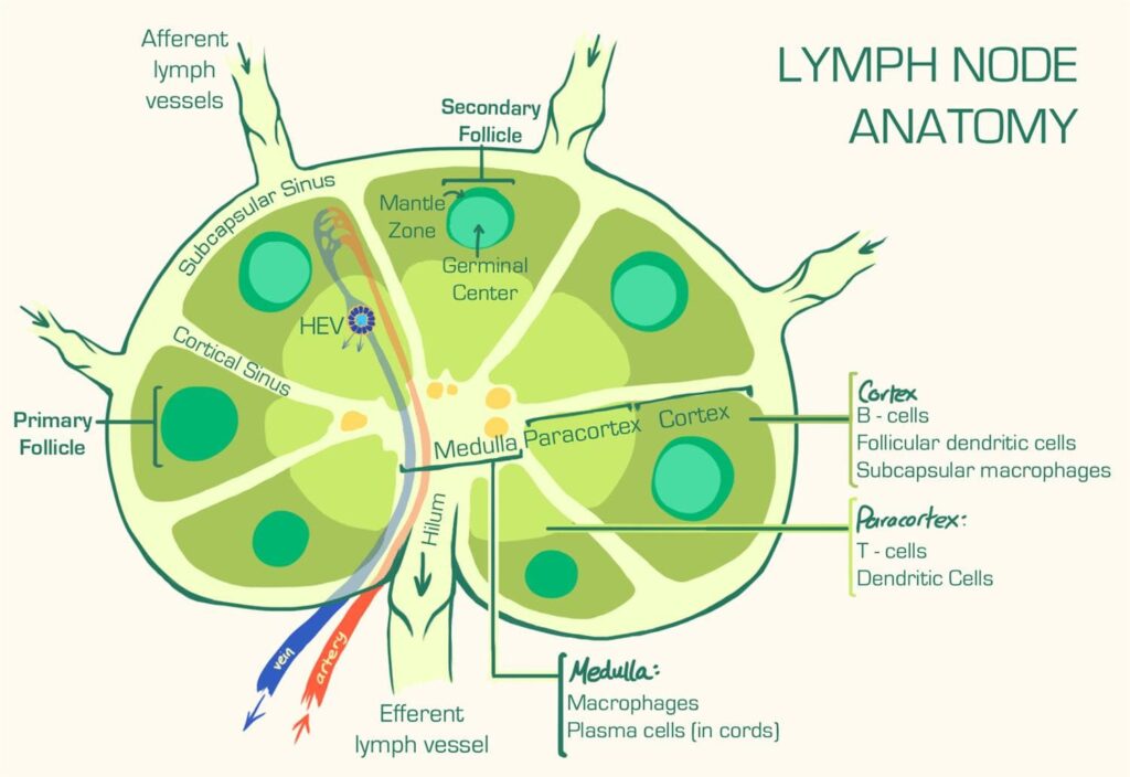
- There are several components of a lymph node, which are divided intothe
- cortex
- an outermost layer of the lymph node that is composed of
- follicles composed of proliferating B cells that can be
- subcapsular macrophages that monitor incoming lymph
- dendritic cells that serve as antigen presenting cells
- an outermost layer of the lymph node that is composed of
- paracortex
- medulla
- an innermost layer of the lymph node that is composed of
- cords with closely packed lymphocyte and plasma cell “cords”
- sinuses that drain into the efferent lymph vessels and house
- reticular cells
- an innermost layer of the lymph node that is composed of
- cortex
- macrophages
Lymph Node Drainage Pattern
| Lymph Node Drainage Pattern | |
| Lymph Node Cluster | Drainage Area |
| Cervical | HeadNeck |
| Hilar | Lungs |
| Mediastinal | TracheaEsophagus |
| Axillary | Upper limbBreastSkin above the umbilicus |
| Celiac | LiverStomachSpleenPancreasUpper duodenum |
| Superior mesenteric | Lower duodenumJejunumIleumColon proximal to splenic flexure |
| Inferior mesenteric | Colon between splenic flexure and upper rectum |
| Internal iliac | Lower rectumAnal canal proximal to pectinate lineBladderCervixProstate |
| Paraaortic | TestesOvaryKidneyUterus |
| Superficial inguinal | Anal canal distal to pectinate lineSkin below the umbilicusScrotumVulva |
| Popliteal | Dorsolateral footPosterior calf |
Secondary lymphoid tissues are vital components of the immune system, serving as hubs for immune cell interactions and the initiation of immune responses. Understanding secondary lymphoid tissues is crucial for medical professionals, especially those in immunology, oncology, and infectious diseases. This article provides a comprehensive overview of secondary lymphoid tissues, including their types, functions, related studies, treatment considerations, and clinical significance.
Types of Secondary Lymphoid Tissues:
- Lymph Nodes: Lymph nodes are small, bean-shaped structures distributed throughout the body, often clustered near major blood vessels. They filter lymphatic fluid, trapping pathogens and antigens for immune cell examination.
- Spleen: The spleen acts as a blood filter, removing damaged blood cells and pathogens. It also contains white pulp, rich in immune cells, where immune responses are initiated.
- Tonsils: Tonsils are collections of lymphoid tissue in the throat and nasal passages. They help prevent infections in the upper respiratory and digestive tracts.
- Peyer’s Patches: These are found in the small intestine and are crucial for monitoring and responding to pathogens in the gut.
- Appendix: The appendix, often considered a vestigial structure, contains lymphoid tissue that may play a role in immune function, particularly in the gut.
- Mucosa-Associated Lymphoid Tissue (MALT): MALT includes various lymphoid structures in mucosal tissues, such as the gut, respiratory tract, and genital tract, providing immunity at mucosal surfaces.
Function of Secondary Lymphoid Tissues:
- Immune Surveillance: Secondary lymphoid tissues act as surveillance centers, where immune cells constantly monitor the body for pathogens and antigens.
- Antigen Presentation: Antigen-presenting cells, such as dendritic cells, macrophages, and B cells, capture antigens in peripheral tissues and migrate to secondary lymphoid tissues to present these antigens to T cells, initiating adaptive immune responses.
- Immune Cell Interaction: Secondary lymphoid tissues facilitate interactions between immune cells, promoting coordinated immune responses.
- Production of Antibodies: B cells within secondary lymphoid tissues generate antibodies in response to antigens, contributing to humoral immunity.
Secondary Lymphoid Tissue Studies:
- Histology and Anatomy: Research focuses on understanding the histological structure and anatomical distribution of secondary lymphoid tissues.
- Immune Cell Trafficking: Studies investigate how immune cells traffic to and from secondary lymphoid tissues during immune responses.
Clinical Significance:
- Infections: Diseases that affect secondary lymphoid tissues can impair immune function and lead to recurrent infections.
- Cancer: Lymphomas and other malignancies often involve lymph nodes and other secondary lymphoid tissues, requiring their evaluation for diagnosis and staging.
Treatment Considerations:
- Infections: In cases of severe or recurrent infections related to secondary lymphoid tissue dysfunction, antimicrobial therapy may be necessary.
- Cancer: For lymphomas and other cancers involving secondary lymphoid tissues, treatment may include chemotherapy, radiation therapy, or surgical removal of affected tissues.
Future Directions:
- Immunotherapies: Ongoing research explores immunotherapies that enhance immune cell function within secondary lymphoid tissues for improved cancer treatment and vaccine development.
- Mucosal Immunization: Investigating the role of MALT in mucosal immunity informs the development of mucosal vaccines and therapies for diseases affecting mucosal surfaces.
Conclusion:
Secondary lymphoid tissues are integral to the immune system, acting as key sites for immune cell interaction, antigen presentation, and the initiation of adaptive immune responses. Lymph nodes, the spleen, tonsils, Peyer’s patches, the appendix, and MALT collectively contribute to immune surveillance and response throughout the body. Understanding their structure, function, and clinical significance is crucial for diagnosing and managing immune-related conditions, including infections and lymphoid tissue-involved cancers.
Check out Interview Preparation, CV and Personal Statement Editing – The Finale.