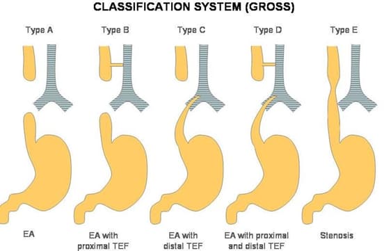
Tracheoesophageal Fistula
- Abnormal connection between esophagus and trachea
- due to blind esophagus, NG tubes cannot be passed into stomach
Tracheoesophageal Fistula (TEF) is a congenital abnormality that involves an abnormal connection between the trachea (windpipe) and the esophagus (food pipe). It is one of the most common congenital anomalies of the respiratory and gastrointestinal systems and requires prompt diagnosis and management. This article provides a comprehensive overview of Tracheoesophageal Fistula, including its types, functions, diagnostic studies, treatment approaches, and prognosis.
Types of Tracheoesophageal Fistula

- Esophageal Atresia (EA) with TEF: This is the most common type of TEF, where a portion of the esophagus is not properly formed, resulting in a blind-ended pouch. The TEF connects the lower part of the trachea to the upper pouch of the esophagus.
- H-Type TEF: In this type, the TEF occurs without esophageal atresia. There is a direct connection between the trachea and the esophagus, without any interruption in the continuity of the esophagus.
Function and Clinical Presentation:
The presence of a Tracheoesophageal Fistula can lead to several significant clinical manifestations:
- Respiratory Distress: Due to the abnormal connection between the trachea and esophagus, food or gastric contents can enter the airway, leading to choking, aspiration, and respiratory distress, especially during feeding.
- Excessive Salivation: Babies with TEF may exhibit excessive drooling because the saliva cannot pass through the esophagus and reach the stomach.
- Cyanosis and Coughing: Infants may present with cyanosis (bluish discoloration of the skin) and coughing during or immediately after feeding due to the aspiration of food into the respiratory tract.
- Abdominal Distension: In cases of EA with TEF, the blind-ended pouch of the esophagus can cause an accumulation of air in the stomach, leading to abdominal distension.
Diagnostic Studies:
- Antenatal Ultrasound: In some cases, TEF can be detected during routine prenatal ultrasound examinations, especially if there are associated anomalies or polyhydramnios (excessive amniotic fluid).
- Chest X-ray: A chest X-ray may reveal the presence of air in the stomach and intestines (pneumoperitoneum) due to the passage of air through the TEF.
- Esophagram (Contrast Study): An esophagram involves administering a contrast medium that is swallowed by the baby. X-rays are then taken to visualize the anatomy of the esophagus and identify the TEF.
- Bronchoscopy: Bronchoscopy is a procedure in which a flexible tube with a camera is inserted into the airways to directly visualize the TEF and assess the tracheobronchial anatomy.
Treatment and Management:
Surgical intervention is the standard treatment for Tracheoesophageal Fistula. The primary goals of surgery are to repair the TEF and, if present, to restore the continuity of the esophagus.
- Preoperative Management: Before surgery, babies with TEF may require suctioning of the airway to prevent aspiration of secretions. Feeding is withheld to avoid further complications.
- Surgical Repair: The surgical repair of TEF involves closing the abnormal connection between the trachea and esophagus and reconstructing the esophagus if there is associated esophageal atresia.
- Gastrostomy Tube: In some cases, a gastrostomy tube (G-tube) may be placed in the stomach to provide nutrition while the repaired esophagus heals.
- Postoperative Care: After surgery, careful monitoring is essential, and infants are gradually introduced to oral feedings once the surgical site has healed.
Prognosis:
The prognosis for infants with TEF depends on the type of TEF, associated anomalies, and the overall health of the baby. With early diagnosis and appropriate surgical intervention, the long-term outcomes are generally favorable. However, there may be potential complications, such as anastomotic leaks (leakage at the surgical site), strictures (narrowing of the esophagus), or gastroesophageal reflux (GERD).
Conclusion:
Tracheoesophageal Fistula (TEF) is a common congenital abnormality involving an abnormal connection between the trachea and the esophagus. It can lead to significant respiratory distress and aspiration of food or gastric contents into the airway. Early diagnosis through antenatal ultrasound or postnatal imaging studies, such as an esophagram, is crucial for timely management.
Surgical repair is the mainstay of treatment, and infants with TEF generally have a good prognosis with appropriate care. Continuous monitoring and follow-up are essential to address any potential complications and ensure the optimal growth and development of affected infants.
Check out USMLE Step 1 Mastery: Comprehensive Course and Lecture Notes.


