Origin of Ulnar Nerve
- Ulnar nerve comes from the medial cord of the brachial plexus (C8-T1)
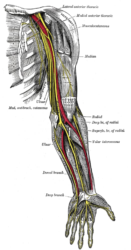
Course of Ulnar Nerve
- Lies posteromedial to brachial artery in anterior compartment of upper 1/2 arm
- Pierces medial IM septa at the arcade of Struthers ~ 8cm from medial epicondyle and lies with triceps
- Travels on back of medial epicondyle; vulnerable in fractures
- Runs with superior ulnar collateral artery
- Cubital tunnel
- roof – cubital tunnel retinaculum (medial epicondyle to olecranon) / osbourne’s fascia (extension of deep forearm fascia between heads of FCU)
- floor – posterior and transverse bands of MCL
- Does send small sensory branch to elbow that can be sacrificed
- Passes into forearm between 2 heads of flexor carpi ulnaris
- Runs between FCU + FDP
- At the wrist, the ulnar nerve and artery pass superficial to the flexor retinaculum
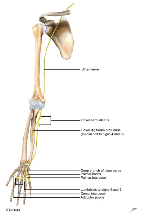
Motor Innervation of Ulnar Nerve
- motor
- forearm
- flexor carpi ulnaris
- flexor digitorum profundus III and IV
- thenar
- adductor pollicis
- deep head of flexor pollicis brevis (FPB)
- fingers
- 3rd & 4th lumbrical (1st & 2nd by median nerve)
- digiti minimi
- abductor digiti minimi
- opponens digiti minimi
- flexor digiti minimi
- forearm
- sensory branches of ulnar nerve
- dorsal cutaneous branch
- palmar cutaneous branch
- superficial terminal branches
Clinical Conditions
- Cubital Tunnel Syndrome compression sites
-
- Arcade of Struthers (tunnel 8 cm proximal to medial epicondyle formed by fibrous connection between IM septum and medial head of triceps)
- medial intermuscular septum
- medial epicondyle (osteophytes)
- cubital tunnel retinaculum (taught with flexion)
- often the retinaculum is consistent with Osborne’s ligament
- aponeurosis of the two heads of the FCU (arcuate ligament) is often consistent with the retinaculum and osbournes ligament, however these fibers meet perpendicular to retinaculum/osbournes ligament
- deep flexor/pronator aponeurosis (most distal site – approximately 4 cm distal to medial epicondyle)
- The internal anatomy of the ulnar n can explain the predominance of hand sx from cubital tunnel syndrome – the fibers to FCU and FDP are central and hand intrinsic fibers are peripheral!
- Ulnar tunnel syndrome: compression in Guyon’s Canal
- no involvement of dorsal cutaneous nerve since it branches before canal
- no involvement of FDP of 4th & 5th and FCU
- ganglia most common cause (from triquetrohamate joint, 32-48%)
- other causes: other mass, trauma (Distal radius/ulna, hook of hamate), muscle anomaly, ulnar artery aneurysm
- Zones of compression
- Zone 1: proximal to bifurcation: hook of hamate fx & ganglia, motor & sensory findings
- Zone 2: deep motor branch; hook of hamate fx & ganglia, motor sx
- Zone 3: superficial sensory branch; ulnar artery thrombosis