

Normal Anatomy (Umbilical Cord and Remnants)
- Umbilical arteries
- right and left umbilical arteries branching from internal iliac arteries return deoxygenated blood from the fetus
- single umbilical artery is associated with increased likelihood of congenital abnormalities
- right and left umbilical arteries branching from internal iliac arteries return deoxygenated blood from the fetus
- Umbilical vein
- single umbilical vein returns oxygenated blood from placenta to fetus
- Allantois
- forms the umbilical arteries and vein
Abnormalities
- Urachal duct
- a duct extending from yolk sac to apex of the bladder
- yolk sac forms allantois which becomes urachus
- during development, the lumen of the uraches closes
- adult remnant is median umbilical ligament
- if urachus fails to close
- patent urachus
- urine leaks from umbilicus
- vesicourachal diverticulum
- bladder protrudes from umbilicus
- patent urachus
- Vitelline duct (omphalomesenteric duct)
- can have ectopic gastric mucosa leading to melena and RLQ pain
Introduction – Umbilical Cord and Remnants
The umbilical cord is a vital structure that connects the developing fetus to the placenta in the womb, allowing essential nutrients and oxygen to be transported from the mother to the baby. After birth, the umbilical cord is clamped and cut, leaving behind a small stump, known as the umbilical remnant or belly button. Understanding the umbilical cord and remnants is crucial for medical professionals, especially those in obstetrics, pediatrics, and neonatology. This article provides a comprehensive overview of the umbilical cord and remnants, including their types, function, related studies, treatment considerations, and clinical significance.
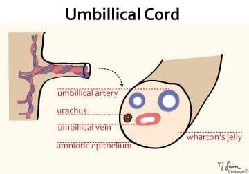
Umbilical Cord Structure:
The umbilical cord is formed during early embryonic development and consists of the following structures:
- Two Umbilical Arteries: These vessels carry deoxygenated blood from the fetus to the placenta for oxygen and nutrient exchange.
- One Umbilical Vein: The umbilical vein carries oxygenated blood and nutrients from the placenta to the fetus.
- Wharton’s Jelly: A gelatinous substance that surrounds and protects the blood vessels, providing structural support to the umbilical cord.
Function of the Umbilical Cord:
The umbilical cord serves as a lifeline for the developing fetus, providing the following functions:
- Nutrient and Oxygen Exchange: Oxygen and essential nutrients, such as glucose and amino acids, are transported from the placenta to the fetus through the umbilical vein.
- Waste Removal: Waste products and carbon dioxide produced by the fetus are carried away from the fetus to the placenta through the umbilical arteries.
Clamping and Cutting of the Umbilical Cord:
After childbirth, the umbilical cord is clamped and cut to separate the baby from the placenta. This procedure is painless and typically performed a few minutes after birth. The cut end of the umbilical cord forms the umbilical remnant.
Umbilical Remnant:
The umbilical remnant, also known as the belly button or navel, is the small stump that remains after the umbilical cord is cut. It gradually dries out and falls off within one to three weeks after birth. The remnant eventually forms the permanent belly button, which is a cosmetic landmark and holds no physiological function after birth.
Types of Umbilical Remnants:
- Normal Healing: In most cases, the umbilical remnant heals without any complications, and the belly button forms as a small scar at the site of the umbilical cord attachment.
- Umbilical Granuloma: Occasionally, a small, reddish, and moist tissue mass called an umbilical granuloma may form at the site of the umbilical remnant due to incomplete healing. This condition is usually harmless and can be treated by a healthcare professional.
Treatment Considerations:
- Umbilical Cord Care: After cutting the umbilical cord, proper care is essential to prevent infection and promote healing. Keeping the area clean and dry is recommended.
- Umbilical Granuloma Treatment: If an umbilical granuloma is present, it can be treated by applying a silver nitrate solution or using other cauterization methods to promote healing.
Clinical Significance:
- Umbilical Cord Abnormalities: Abnormalities in the umbilical cord, such as a single umbilical artery or nuchal cord (cord wrapped around the baby’s neck), can be identified through prenatal ultrasound and may require additional monitoring during pregnancy.
- Umbilical Remnant Infections: Occasionally, the umbilical remnant may become infected, leading to signs of redness, swelling, and discharge. Prompt medical attention and appropriate treatment are necessary in such cases.
Umbilical Cord Studies:
- Prenatal Ultrasound: Prenatal ultrasound is a valuable tool for evaluating the umbilical cord and identifying any abnormalities during pregnancy.
Conclusion:
The umbilical cord is a crucial structure that provides essential nutrient and oxygen exchange between the fetus and placenta during pregnancy. After childbirth, the umbilical cord is clamped and cut, leaving behind the umbilical remnant, which eventually forms the belly button. Proper umbilical cord care and monitoring of the umbilical remnant are essential for preventing infections and promoting healing. In cases of abnormal umbilical cord findings during prenatal ultrasound, additional monitoring and management may be required to ensure a healthy pregnancy and delivery. Overall, understanding the umbilical cord and remnants is vital for providing optimal care to newborns and their families during the perinatal period.
Check out USMLE Step 1 Mastery: Comprehensive Course and Lecture Notes.